Spectrum of Cervical Lesions Among Women Undergoing Cervical Biopsy: A Cross-Sectional Study
DOI:
https://doi.org/10.68041/jbpt.v2i3/06Keywords:
Biopsy, Postmenopause, Uterine Cervical Diseases, Uterine Cervical Dysplasia, Uterine Cervical Neoplasms, Uterine CervicitisAbstract
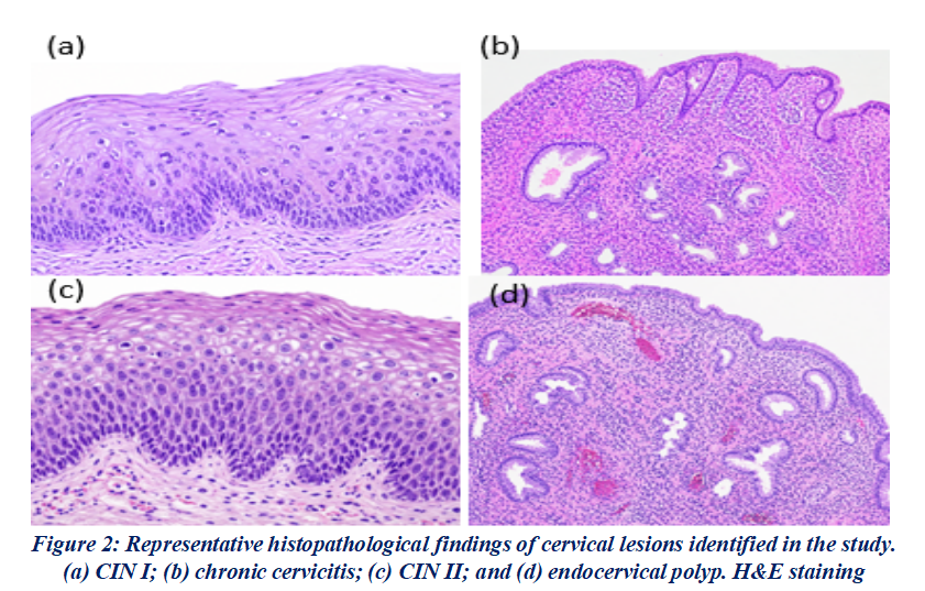
Background: The standard method of determining benign, pre-cancerous, and cancerous cervical lesions is histopathology of biopsy tissue from the cervix. This study aimed to identify the histopathological spectrum of lesions encountered in women who had undergone cervical biopsy and to evaluate the relationship between the lesions and menopausal status. Methods: A retrospective descriptive study design was used to analyze 186 cervical biopsy specimens obtained over the last two years at a tertiary care center. Pathology records were used to obtain demographic and clinical information, including age, indication for biopsy, menopausal status, and histopathological diagnosis. The lesions were graded as benign, premalignant (CIN I-III), and malignant. Association was tested by the Chi-square test and analyzed using SPSS version 26.0 with p<0.05 as significant. Results: The mean age was 43.8 ± 11.6 years. The most common indication for a biopsy was abnormal vaginal bleeding 58(31.2%). Chronic cervicitis was the most common single diagnosis 55(29.6%), followed by CIN I 13 (19.9%) and CIN II 28(15.1%). The overall distribution of premalignant lesions, benign lesions, and malignant lesions was 84(45.2%), 23(37.1%), and 33(17.7%), respectively. Squamous cell carcinoma was the most common form of cancer. Post menopausal women had significantly more malignant lesions than premenopausal women (23(37.1%) vs 10(8.1%), p<0.001). Conclusion: The largest histopathological group was the premalignant lesions, and the commonest individual lesion was chronic cervicitis, indicating that early biopsy diagnosis and robust cervical screening programme, especially for postmenopausal women, is warranted. Future studies considering HPV status can enhance risk stratification and follow-up.
References
1. Basoya S, Anjankar A. Cervical cancer: early detection and prevention in reproductive age group. Cureus. 2022;14(11):e31312. https://doi.org/10.7759/cureus.31312 DOI: https://doi.org/10.7759/cureus.31312
2. Li Z, Liu P, Yin A, Zhang B, Xu J, Chen Z, et al. Global landscape of cervical cancer incidence and mortality in 2022 and predictions to 2030: The urgent need to address inequalities in cervical cancer. Int J Cancer. 2025;157(2):288-297. https://doi.org/10.1002/ijc.35369 DOI: https://doi.org/10.1002/ijc.35369
3. Xu HH, Wang K, Feng XJ, Dong SS, Lin A, Zheng LZ, et al. Prevalence of human papillomavirus genotypes and relative risk of cervical cancer in China: A systematic review and meta-analysis. Oncotarget. 2018;9(20):15386-15397. https://doi.org/10.18632/oncotarget.24169 DOI: https://doi.org/10.18632/oncotarget.24169
4. Cangelosi G, Sacchini F, Mancin S, Petrelli F, Amendola A, Fappani C, et al. Papillomavirus vaccination programs and knowledge gaps as barriers to implementation: A systematic review. Vaccines (Basel). 2025;13(5):460. https://doi.org/10.3390/vaccines13050460 DOI: https://doi.org/10.3390/vaccines13050460
5. Balan TA, Balan RA, Socolov D, Gheorghiță VR, Buțureanu TA, Păvăleanu I, et al. Pregnancy-related precancerous cervical lesions: Pathogenesis, diagnosis, evolution, and impact upon gestation and fertility. J Clin Med. 2024;13(22):6718. https://doi.org/10.3390/jcm13226718 DOI: https://doi.org/10.3390/jcm13226718
6. Bruno MT, Pagana A, Lo Giudice C, Panella MM, Mascellino G, Laganà AS. CIN2 in the era of risk-based management and HPV vaccination: Epidemiology, natural history and guidelines. Diagnostics (Basel). 2025;15(19):2512. https://doi.org/10.3390/diagnostics15192512 DOI: https://doi.org/10.3390/diagnostics15192512
7. Gupta S, Nagtode N, Chandra V, Gomase K. From diagnosis to treatment: Exploring the latest management trends in cervical intraepithelial neoplasia. Cureus. 2023;15(12):e50291. https://doi.org/10.7759/cureus.50291 DOI: https://doi.org/10.7759/cureus.50291
8. Jadhav A, Sanklecha V, Natekar A, Mahra R. Histopathological study of spectrum of lesions of uterine cervix. J Midlife Health. 2023;14(2):112-116. https://doi.org/10.4103/jmh.jmh_28_23 DOI: https://doi.org/10.4103/jmh.jmh_28_23
9. Sozen I, Sahin G, Ulu Y, Yitiz D, Kayan BO, Yuksel IT. Cytology-biopsy concordance in high-risk human papillomavirus-positive women with abnormal cytology findings: Menopause-stratified analysis. Medicina (Kaunas). 2026;62(4):631. https://doi.org/10.3390/medicina62040631 DOI: https://doi.org/10.3390/medicina62040631
10. Gisca T, Matasariu DR, Ursache A, Socolov DG, Scripcariu IS, Fudulu A, et al. Integrating biomarkers into cervical cancer screening—Advances in diagnosis and risk prediction: A narrative review. Diagnostics (Basel). 2025;15(24):3231. https://doi.org/10.3390/diagnostics15243231 DOI: https://doi.org/10.3390/diagnostics15243231
11. Sullivan KM, Dean A, Soe MM. OpenEpi: a web-based epidemiologic and statistical calculator for public health. Public Health Rep. 2009;124(3):471–474. https://doi.org/10.1177/003335490912400320 DOI: https://doi.org/10.1177/003335490912400320
12. Goodyear MDE, Krleza-Jeric K, Lemmens T. The Declaration of Helsinki. BMJ. 2007;335(7621):624–625. https://doi.org/10.1136/bmj.39339.610000.BE DOI: https://doi.org/10.1136/bmj.39339.610000.BE
13. Kaseka PU, Kayira A, Chimbatata CS, Chisale MRO, Kamudumuli P, Wu TSJ, et al. Histopathological profile of cervical biopsies in northern Malawi: A retrospective cross-sectional study. BMJ Open. 2022;12(3):e048283. https://doi.org/10.1136/bmjopen-2020-048283 DOI: https://doi.org/10.1136/bmjopen-2020-048283
14. Kerthi KS, Chander V. A study of histopathological spectrum of lesions in cervix biopsies in a tertiary care hospital. J Pharm Res Int. 2021;33(64B):47-53. https://doi.org/10.9734/jpri/2021/v33i64B35330 DOI: https://doi.org/10.9734/jpri/2021/v33i64B35330
15. Deepika, Khan A, Salmani A. Histopathological spectrum of cervical lesions in women attending a tertiary care hospital. International Journal of Clinical Obstetrics and Gynaecology. 2026;10(2):833–835. https://doi.org/10.33545/gynae.2026.v10.i2k.2127 DOI: https://doi.org/10.33545/gynae.2026.v10.i2k.2127
16. Loopik DL, Bentley HA, Eijgenraam MN, IntHout J, Bekkers RLM, Bentley JR. The natural history of cervical intraepithelial neoplasia grades 1, 2, and 3: a systematic review and meta-analysis. Journal of Lower Genital Tract Disease. 2021;25(3):221–231. https://doi.org/10.1097/LGT.0000000000000604 DOI: https://doi.org/10.1097/LGT.0000000000000604
17. Gahine R, Pandey V, Gajendra M, Shende PK. Analysis of pattern of expression of immunohistochemical markers p16, p53 and Ki67 on cervical lesions and its correlation with histopathology. Journal of Medical Sciences and Health. 2025;11(1):32–41. https://doi.org/10.46347/jmsh.v11.i1.24.242 DOI: https://doi.org/10.46347/jmsh.v11.i1.24.242
18. Sakai M, Ohara T, Suzuki H, Kadomoto T, Inayama Y, Shitanaka S, et al. Clinical impact of age-specific distribution of combination patterns of cytology and high-risk HPV status on cervical intraepithelial neoplasia grade 2 or more. Oncology Letters. 2023;26(3):384. https://doi.org/10.3892/ol.2023.13970 DOI: https://doi.org/10.3892/ol.2023.13970
19. Rasool S, Naz F, Abdul Wahab T, Khidri FF, Riaz H, Abbasi N. Frequency of abnormal Pap smear in patients presenting with postcoital bleeding in tertiary care hospital, Karachi, Pakistan. Tropical Doctor. 2023;53(2):267–270. https://doi.org/10.1177/00494755221151092 DOI: https://doi.org/10.1177/00494755221151092
20. Gothwal M, Singh P, Elhence P, Yadav G, Sharma C, Samriya N. Spectrum of abnormal cervical lesions and its cytohistological correlation: a study from an institute of national importance of Western Rajasthan. Maedica (Bucur). 2022;17(3):636–640. https://doi.org/10.26574/maedica.2022.17.3.636 DOI: https://doi.org/10.26574/maedica.2022.17.3.636
21. Choi MS, Lee YJ, Lee EH, Ji YI, Park MJ. Factors associated with cyto-histological misinterpretation of cervical smear according to menopausal status. Journal of Menopausal Medicine. 2022;28(2):78–84. https://doi.org/10.6118/jmm.22023 DOI: https://doi.org/10.6118/jmm.22023
22. Jia M, Lan C, Niu J, Liang Y. Risk factors for pathological upgrading in perimenopausal women with cervical intraepithelial neoplasia grade 2/3 following conization. Medicine (Baltimore). 2022;101(43):e31368. https://doi.org/10.1097/MD.0000000000031368 DOI: https://doi.org/10.1097/MD.0000000000031368
23. Salibay C, Chen Z, Ma B, Pan H, Hijazi M, Elatre W, et al. High-risk HPV testing improves accuracy in detection of CIN2+ lesions in ASC-H postmenopausal women? An academic hospital experience. J Am Soc Cytopathol. 2023;12(1):58–65. doi: https://doi.org/10.1016/j.jasc.2022.08.004 DOI: https://doi.org/10.1016/j.jasc.2022.08.004
24. Ouh YT, Kim HY, Yi KW, Lee NW, Kim HJ, Min KJ. Enhancing cervical cancer screening: Review of p16/Ki-67 dual staining as a promising triage strategy. Diagnostics (Basel). 2024;14(4):451. https://doi.org/10.3390/diagnostics14040451 DOI: https://doi.org/10.3390/diagnostics14040451
Published
Issue
Section
License
Copyright (c) 2026 Aliya Iftikhar Memon, Rukhsana Izhar, Zarrien Fatima, Ayesha Kamran

This work is licensed under a Creative Commons Attribution-NonCommercial 4.0 International License.
Journal of Biomolecules, Pathogenesis and Therapeutics (JBPT) follows the Attribution Creative Commons-Non commercial (CC BY-NC 4.0) license which allows the users to copy and redistribute the material in any medium or format, remix, transform and build upon the material. The users must give credit to the source and indicate, provide a link to the license, and indicate if changes were made. However, the CC BY-NC 4.0 license restricts the use of material for commercial purposes (Further details are available at website. The Editorial Board of the JBPT endeavors to ensure the accuracy, integrity, and quality of all published content. However, the responsibility of opinions, interpretations, and conclusions expressed in the published articles rests entirely with the authors and do not necessarily reflect the views of the Editorial Board, publisher, or affiliated institutions.