Expression profiling of TP53 and diagnostic utility in Breast Cancer: clinical and molecular Perspectives
DOI:
https://doi.org/10.68041/jbpt.v2i2/05Keywords:
Breast cancer, TP53, gene expression, RT-PCR, biomarker, molecular diagnostics, tumor suppressor geneAbstract
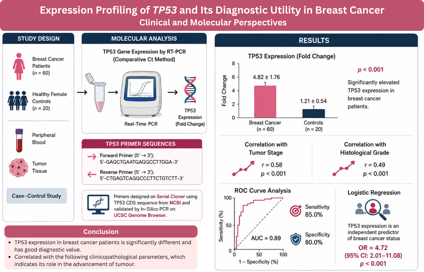
Background: Breast cancer is the most prevalent malignancy in women worldwide and remains a major cause of deaths due to cancer. This study measured TP53 gene expression in patients with breast cancer and to determine its possible diagnostic value in breast cancer patients by correlating the TP53 gene expression with clinical and pathological parameters. Methodology: This was a case-control study with 80 participants; 60 patients with histopathologically confirmed breast cancer and 20 healthy female controls. Peripheral blood and tumor tissue specimens were taken for molecular studies. The TP53 gene expression level was measured by real-time polymerase chain reaction (RT-PCR) using comparative Ct method. Statistical analysis was carried out by SPSS version 22.0 (p < 0.05 level). Results: The TP53 expression was considerably higher in breast cancer patients, by comparing healthy controls (4.82 ± 1.76 vs. 1.21 ± 0.54 fold change, p < 0.001). Both tumor stage (r = 0.58, p < 0.001) and histological grade (r = 0.49, p < 0.001) were positively interrelated with TP53 expression. The ROC curve analysis showed an area under the curve (AUC) of 0.89, sensitivity of 85.0% and specificity of 80.0%. The expression of TP53 was an independent predictor of breast cancer status identified by logistic regression (OR = 4.72, 95% CI: 2.01–11.08, p < 0.001). Conclusion: TP53 expression in breast cancer patients is significantly different and has good diagnostic value. It is correlated with the following clinicopathological parameters, which indicates its role in the advancement of tumor.
References
1. Łukasiewicz S, Czeczelewski M, Forma A, Baj J, Sitarz R, Stanisławek A. Breast Cancer-Epidemiology, Risk Factors, Classification, Prognostic Markers, and Current Treatment Strategies-An Updated Review. Cancers (Basel). 2021 Aug 25;13(17):4287. https://doi.org/10.3390/cancers13174287
2. Zhao N, Rosen JM. Breast cancer heterogeneity through the lens of single-cell analysis and spatial pathologies. Semin Cancer Biol. 2022 Jul;82:3-10. https://doi.org/10.1016/j.semcancer.2021.07.010
3. Lee AC, Lee HB, Yeom H, Song SW, Kim SD, Choi A, et al. Advances in Tumor Sampling and Sequencing in Breast Cancer and their Application in Precision Diagnostics and Therapeutics. Adv Exp Med Biol. 2021;1187:215-244. https://doi.org/10.1007/978-981-32-9620-6_11
4. Yoo TK. Liquid Biopsy in Breast Cancer: Circulating Tumor Cells and Circulating Tumor DNA. Adv Exp Med Biol. 2021;1187:337-361. https://doi.org/10.1007/978-981-32-9620-6_17
5. Chen X, Guo Y, Ouyang T, Li J, Wang T, Fan Z, et al. Co-mutation of TP53 and PIK3CA in residual disease after neoadjuvant chemotherapy is associated with poor survival in breast cancer. J Cancer Res Clin Oncol. 2019 May;145(5):1235-1242. https://doi.org/10.1007/s00432-019-02873-8
6. Qayoom H, Haq BU, Sofi S, Jan N, Jan A, Mir MA. Targeting mutant p53: a key player in breast cancer pathogenesis and beyond. Cell Commun Signal. 2024 Oct 10;22(1):484. https://doi.org/10.1186/s12964-024-01863-9
7. Anderson SA, Bartow BB, Harada S, Siegal GP, Wei S, Dal Zotto VL, et al. p53 protein expression patterns associated with TP53 mutations in breast carcinoma. Breast Cancer Res Treat. 2024 Aug;207(1):213-222. https://doi.org/10.1007/s10549-024-07357-z
8. Lee H, Cho YA, Kim DG, Son JY, Cho EY. Real-world Comparison of P53 Immunohistochemistry and TP53 Mutation Analysis Using Next-generation Sequencing. Anticancer Res. 2024 Sep;44(9):3983-3994. https://doi.org/10.21873/anticanres.17227
9. Budhbaware T, Rathored J, Shende S. Molecular methods in cancer diagnostics: a short review. Ann Med. 2024 Dec;56(1):2353893. https://doi.org/10.1080/07853890.2024.2353893
10. Chen X, Guo Y, Ouyang T, Li J, Wang T, Fan Z, et al. Co-mutation of TP53 and PIK3CA in residual disease after neoadjuvant chemotherapy is associated with poor survival in breast cancer. J Cancer Res Clin Oncol. 2019 May;145(5):1235-1242. https://doi.org/10.1007/s00432-019-02873-8
11. Shahbandi A, Nguyen HD, Jackson JG. TP53 Mutations and Outcomes in Breast Cancer: Reading beyond the Headlines. Trends Cancer. 2020 Feb;6(2):98-110. https://doi.org/10.1016/j.trecan.2020.01.007
12. Mahtani R, Holmes FA, Badve S, Caldera H, Coleman R, Mamounas E, et al. Breast Cancer Therapy Expert Group (BCTEG). A Roundtable Discussion of the Breast Cancer Therapy Expert Group (BCTEG): Clinical Developments and Practice Guidance on Human Epidermal Growth Factor Receptor 2 (HER2)-positive Breast Cancer. Clin Breast Cancer. 2020 Jun;20(3):e251-e260. https://doi.org/10.1016/j.clbc.2019.08.001
13. Mueller S, Grote I, Bartels S, Kandt L, Christgen H, Lehmann U, et al. p53 Expression in Luminal Breast Cancer Correlates With TP53 Mutation and Primary Endocrine Resistance. Mod Pathol. 2023 Apr;36(4):100100. https://doi.org/10.1016/j.modpat.2023.100100
14. Arab D, Fakhar HBZ, Rouhollah F, Akbari ME, Jalalian M, Shahidi PS. Analysis of tumor protein p53 expression levels in different stages of breast cancer. Sci Rep. 2025 Nov 13;15(1):39894. https://doi.org/10.1038/s41598-025-23713-5
15. Manoharan V, Karunanayake EH, Tennekoon KH, De Silva S, Imthikab AIA, De Silva K, et al. Pattern of nucleotide variants of TP53 and their correlation with the expression of p53 and its downstream proteins in a Sri Lankan cohort of breast and colorectal cancer patients. BMC Cancer. 2020 Jan 30;20(1):72. https://doi.org/10.1186/s12885-020-6573-5
16. Huang Y, Ren S, Ding L, Jiang Y, Luo J, Huang J, et al. TP53-specific mutations serve as a potential biomarker for homologous recombination deficiency in breast cancer: a clinical next-generation sequencing study. Precis Clin Med. 2024 Apr 9;7(2):pbae009. https://doi.org/10.1093/pcmedi/pbae009
17. Shahbandi A, Nguyen HD, Jackson JG. TP53 Mutations and Outcomes in Breast Cancer: Reading beyond the Headlines. Trends Cancer. 2020 Feb;6(2):98-110. https://doi.org/10.1016/j.trecan.2020.01.007
18. Tang Q, Su Z, Gu W, Rustgi AK. Mutant p53 on the Path to Metastasis. Trends Cancer. 2020 Jan;6(1):62-73. https://doi.org/10.1016/j.trecan.2019.11.004
19. Darb-Esfahani S, Denkert C, Stenzinger A, Salat C, Sinn B, Schem C, et al. Role of TP53 mutations in triple negative and HER2-positive breast cancer treated with neoadjuvant anthracycline/taxane-based chemotherapy. Oncotarget. 2016 Oct 18;7(42):67686-67698. https://doi.org/10.18632/oncotarget.11891
20. Steffens Reinhardt L, Groen K, Morten BC, Bourdon JC, Avery-Kiejda KA. Cytoplasmic p53β Isoforms Are Associated with Worse Disease-Free Survival in Breast Cancer. Int J Mol Sci. 2022 Jun 15;23(12):6670. https://doi.org/10.3390/ijms23126670
21. Steffens Reinhardt L, Groen K, Xavier A, Avery-Kiejda KA. p53 Dysregulation in Breast Cancer: Insights on Mutations in the TP53 Network and p53 Isoform Expression. Int J Mol Sci. 2023 Jun 13;24(12):10078. https://doi.org/10.3390/ijms241210078
22. Marvalim C, Datta A, Lee SC. Role of p53 in breast cancer progression: An insight into p53 targeted therapy. Theranostics. 2023 Feb 27;13(4):1421-1442. https://doi.org/10.7150/thno.81847
23. Darb-Esfahani S, Denkert C, Stenzinger A, Salat C, Sinn B, Schem C, et al. Role of TP53 mutations in triple negative and HER2-positive breast cancer treated with neoadjuvant anthracycline/taxane-based chemotherapy. Oncotarget. 2016 Oct 18;7(42):67686-67698. https://doi.org/10.18632/oncotarget.11891
24. Xiong M, Wang X, Liu D, Xiu B, Zhang Q, Chi W, et al. Somatic mutations in a multigene panel and impact on prognosis based on TP53 status in Chinese HER2-positive patients undergoing neoadjuvant therapy: A single-institution retrospective cohort. Cancer Med. 2024 Jan;13(2):e6955. https://doi.org/10.1002/cam4.6955
Published
Issue
Section
License
Copyright (c) 2026 Maryum Qayyum, Kashif Shahbaz, Khadija Ahmed, Mubeen Anjum (Author)

This work is licensed under a Creative Commons Attribution-NonCommercial 4.0 International License.
Journal of Biomolecules, Pathogenesis and Therapeutics (JBPT) follows the Attribution Creative Commons-Non commercial (CC BY-NC 4.0) license which allows the users to copy and redistribute the material in any medium or format, remix, transform and build upon the material. The users must give credit to the source and indicate, provide a link to the license, and indicate if changes were made. However, the CC BY-NC 4.0 license restricts the use of material for commercial purposes (Further details are available at website. The Editorial Board of the JBPT endeavors to ensure the accuracy, integrity, and quality of all published content. However, the responsibility of opinions, interpretations, and conclusions expressed in the published articles rests entirely with the authors and do not necessarily reflect the views of the Editorial Board, publisher, or affiliated institutions.