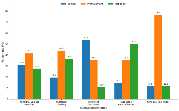
Figure 1: Distribution of benign, premalignant, and malignant histopathological findings according to clinical presentation
Aliya Iftikhar Memon1 | Rukhsana Izhar1 | Zarrien Fatima2* | Ayesha Kamran3
1Department of Pathology, AIMS, Karachi, Pakistan | 2Department of Medical Microbiology, Quaid-i-Azam University Islamabad, Pakistan
3Department of Molecular Biology, Superior University Lahore, Pakistan
*Correspondence: Zarrien Fatima ([email protected])
Received: 07 August, 2026; Revised: 05 September, 2026; Accepted: 12 September, 2026; Published: 20 September, 2026
Background: The standard method of determining benign, pre-cancerous, and cancerous cervical lesions is histopathology of biopsy tissue from the cervix. This study aimed to identify the histopathological spectrum of lesions encountered in women who had undergone cervical biopsy and to evaluate the relationship between the lesions and menopausal status. Methods: A retrospective descriptive study design was used to analyze 186 cervical biopsy specimens obtained over the last two years at a tertiary care center. Pathology records were used to obtain demographic and clinical information, including age, indication for biopsy, menopausal status, and histopathological diagnosis. The lesions were graded as benign, premalignant (CIN I-III), and malignant. Association was tested by the Chi-square test and analyzed using SPSS version 26.0 with p<0.05 as significant. Results: The mean age was 43.8 ± 11.6 years. The most common indication for a biopsy was abnormal vaginal bleeding 58(31.2%). Chronic cervicitis was the most common single diagnosis 55(29.6%), followed by CIN I 13 (19.9%) and CIN II 28(15.1%). The overall distribution of premalignant lesions, benign lesions, and malignant lesions was 84(45.2%), 23(37.1%), and 33(17.7%), respectively. Squamous cell carcinoma was the most common form of cancer. Post menopausal women had significantly more malignant lesions than premenopausal women (23(37.1%) vs 10(8.1%), p<0.001). Conclusions: The largest histopathological group was the premalignant lesions, and the commonest individual lesion was chronic cervicitis, indicating that early biopsy diagnosis and robust cervical screening programme, especially for postmenopausal women, is warranted. Future studies considering HPV status can enhance risk stratification and follow-up.
Keywords: Biopsy; Postmenopause; Uterine Cervical Diseases; Uterine Cervical Dysplasia; Uterine Cervical Neoplasms; Uterine Cervicitis.
Cervical cancer is still one of the most common types of cancers worldwide despite its being treatable and largely preventable with human papillomavirus (HPV) vaccination, screening, and treatment of precursor lesions 1. The analyses conducted based on the data from GLOBOCAN 2022 estimated that around 662,000 new cases and 349,000 deaths would occur worldwide, and this burden would be disproportionately borne by the less well-resourced areas 2. Regional meta-analysis shows the high prevalence of HPV among women with cervical cancer, and HPV is the major etiological factor in cervical carcinogenesis 3. Despite this, HPV vaccine uptake is low, and there are still significant gaps in organized screening, as shown in evidence from South Asia 4.
The diseases of the cervix can be benign inflammatory and reactive lesions or cervical intraepithelial neoplasia (CIN) and invasive carcinoma 5. Systematic-review evidence indicates that many CIN1 and CIN2 lesions resolve, while progression and persistence of increasingly higher histological grade is a cause for concern 6. Clinically, it is important to differentiate between these categories because the management is different for each, and accurate distinction between these categories is of paramount importance 7. Therefore, the reference standard for confirmation of disease and grading of cervical precancer is histopathology of the uterine cervical tissue 8. The diagnostic usefulness of colposcopy-directed biopsy, however, is affected by age, characteristics of the transformation zone, and menopausal status, and cytological-histological discrepancies are more likely to be a problem post-menopause 9. However, local histopathological data are crucial, even in view of the progress in HPV-based screening, as it has been observed that the distribution of these benign, pre-cancerous and cancerous lesions depends on the characteristics of the population, referral patterns and screening rates. A clear definition of this spectrum can aid in the diagnostic process, in risk stratification, and in planning for cervical-cancer prevention 10. This study will provide locally relevant evidence to support accurate diagnosis and appropriate management of cervical lesions. The present study aimed to identify the histopathological spectrum of cervical lesions in women who underwent cervical biopsy. It was also designed to identify whether lesions were benign, pre-cancerous, or cancerous. The association of histopathological category with menopausal status was also assessed.
A retrospective descriptive study was conducted in a tertiary care teaching hospital in the Histopathology Department. During a 24-month period, 186 consecutive cervical biopsy specimens were included in the study. Sample size was calculated using OpenEpi, Version 3.03 (Emory University, Atlanta, GA, USA; RRID:SCR_021913) based on the expected prevalence of cervical lesions, a 95% confidence level, and an absolute precision of 5%. 11. The sample size was calculated using the formula n = Z2(1-P)/e2, where n represents the required sample size, Z is the standard normal deviate corresponding to the confidence level, P is the expected prevalence, and e is the absolute precision. Cases were identified from the departmental pathology database, and the histopathology reports and request forms were analysed.
All punch biopsies, loop electrosurgical excision procedure (LEEP) specimens and cervical tissue biopsies with a full histopathological report were included. Samples with poor quality tissue, samples with inconclusive diagnosis, duplicate records, and samples with clinical information that was lacking were not analysed. Patient demographic information on the request form for the pathologist included the age and menopausal status of each patient, and clinical indications for biopsy. Histopathological diagnosis was made using the routine hematoxylin and eosin-stained sections prepared using the standard laboratory technique. All the tissue samples were fixed in 10% neutral buffered formalin, processed in graded alcohol and xylene, embedded in paraffin wax, sectioned at approximately 4-5 microns and stained with H&E. All the slides were reviewed and reported by the consultants' histopathologists following standard diagnostic criteria.
Histopathological results were classified as benign lesions, low-grade squamous intraepithelial lesions (LSIL/CIN 1), high-grade squamous intraepithelial lesions (HSIL/CIN 2 and CIN 3), and invasive malignancies. In addition, indications for biopsy were divided into clinical categories for comparison. The data obtained were tabulated and analysed through the use of Statistical Package for Social Sciences IBM SPSS Statistics, Version 28.0 (Released 2021; IBM Corp., Armonk, NY, USA; RRID:SCR_016479). For continuous variables, means ± standard deviations (SDs) were reported, and for categorical variables, frequencies and percentages were reported. The Chi-square test was employed to evaluate the association between menopausal status and histopathological categories, and a p-value < 0.05 was considered statistically significant. The Institutional Review Board has approved the study before data collection. Patients were anonymised during data extraction, and the study uses the ethical principles of the Declaration of Helsinki 12.
A total of 186 specimens of cervical biopsy were included in the histopathological review. The mean age of the patients was 43.8 ± 11.6 years (range: 22–74 years). The most frequent histopathological diagnosis was chronic cervicitis 55(29.6%) followed by cervical intraepithelial neoplasia (CIN) grade I (19.9%) and CIN grade II 28(15.1%). Malignant lesions were the next most common biopsy type 33(17.7%), with the predominant invasive malignancy being squamous cell. As shown in Table I, abnormal vaginal bleeding 58(31.2%), postcoital bleeding 41 (22.0%), and clinically suspicious cervical lesions 34(18.3%) were the most common indications of cervical biopsy. Abnormal cervical cytology was the most common indication for women to be referred for histological evaluation of their cervix, accounting for 17(9.1%) of all women referred for biopsy; screening plays a pivotal role in identifying women who require referral for histological evaluation of the cervix.
| Clinical Indication | Frequency (n) | Percentage (%) |
|---|---|---|
| Abnormal vaginal bleeding | 58 | 31.2 |
| Postcoital bleeding | 41 | 22.0 |
| Suspicious cervical lesion | 34 | 18.3 |
| Persistent vaginal discharge | 28 | 15.1 |
| Abnormal Pap smear | 17 | 9.1 |
| Other indications | 8 | 4.3 |
Chronic cervicitis was the most frequent diagnosis, accounting for 55(29.6%) specimens, followed by CIN I 37(19.9%), CIN II 28(15.1%), CIN III 19(10.2%), squamous cell carcinoma 24(12.9%), adenocarcinoma 9(4.8%), endocervical polyp 8(4.3%), and other benign lesions 6 (3.2%). Overall, CIN I–III together accounted for 84(45.2%) specimens, while benign and malignant lesions accounted for 69(37.1%) and 33(17.7%), respectively, as shown in Table II.
| Histopathological diagnosis | n | % |
|---|---|---|
| Chronic cervicitis | 55 | 29.6 |
| CIN I | 37 | 19.9 |
| CIN II | 28 | 15.1 |
| CIN III | 19 | 10.2 |
| Squamous cell carcinoma | 24 | 12.9 |
| Adenocarcinoma | 9 | 4.8 |
| Endocervical polyp | 8 | 4.3 |
| Other benign lesions | 6 | 3.2 |
| Total | 186 | 100.0 |
The pre-malignant lesions were more prevalent among females aged 30-49 years, while malignant lesions tended to be more common with age. The age group with the highest percentage of women presenting with invasive cervical pathology was the elderly group; almost half of the women in the age group of 60 years and above had malignant histopathological results, as presented in Table III.
| Age Group (years) | Benign n (%) | Premalignant (CIN I–III) n (%) | Malignant n (%) | Total n (%) |
|---|---|---|---|---|
| 20–29 | 16 (23.2%) | 7 (8.3%) | 0 (0.0%) | 23 (12.4%) |
| 30–39 | 23 (33.3%) | 25 (29.8%) | 3 (9.1%) | 51 (27.4%) |
| 40–49 | 17 (24.6%) | 28 (33.3%) | 10 (30.3%) | 55 (29.6%) |
| 50–59 | 10 (14.5%) | 16 (19.0%) | 11 (33.3%) | 37 (19.9%) |
| ≥60 | 3 (4.3%) | 8 (9.5%) | 9 (27.3%) | 20 (10.8%) |
| Total | 69 (100.0%) | 84 (100.0%) | 33 (100.0%) | 186 (100.0%) |
The largest number of malignant diagnoses 17(50.0%) was obtained from women who had suspicious cervical lesions. However, abnormal Pap smear were primarily associated with precancerous cervical changes 13(76.4%), proving the value of cervical cytology in the identification of abnormal epithelial changes early in the disease process, as presented in Table IV.
| Clinical Presentation | Benign n (%) | Premalignant n (%) | Malignant n (%) |
|---|---|---|---|
| Abnormal vaginal bleeding | 18 (31.0) | 24 (41.4) | 16 (27.6) |
| Postcoital bleeding | 8 (19.5) | 18 (43.9) | 15 (36.6) |
| Persistent discharge | 15 (53.6) | 10 (35.7) | 3 (10.7) |
| Suspicious cervical lesion | 5 (14.7) | 12 (35.3) | 17 (50.0) |
| Abnormal Pap smear | 2 (11.8) | 13 (76.4) | 2 (11.8) |
Figure 1 illustrates the distribution of benign, premalignant, and malignant histopathological findings according to the clinical presentation leading to cervical biopsy. Premalignant lesions were particularly frequent among women referred because of abnormal Pap smear findings. Malignant lesions were most frequent among women presenting with clinically suspicious cervical lesions, whereas benign lesions were more commonly observed among those with persistent vaginal discharge.
Figure 1: Distribution of benign, premalignant, and malignant histopathological findings according to clinical presentation
The most common histopathology overall was premalignant lesions 84 (45.2%), benign 69 (37.1%), and malignant 33 (17.7%). There were 54(43.5%) of women with benign lesions, 60 (48.4%) with premalignant lesions, and 8.1% with malignant lesions, among premenopausal women. Malignant lesions, on the other hand, were significantly more common in the postmenopausal women 23 (37.1%) than in the premenopausal women 10 (8.1%). There was a significant association between menopausal status and histopathological category (p<0.001), as shown in Table V.
| Histopathological Category | Premenopausal (n=124), n (%) | Postmenopausal (n=62), n (%) | Total (N=186), n (%) | p-value |
|---|---|---|---|---|
| Benign lesions | 54 (43.5) | 15 (24.2) | 69 (37.1) | |
| Premalignant lesions | 60 (48.4) | 24 (38.7) | 84 (45.2) | <0.001 |
| Malignant lesions | 10 (8.1) | 23 (37.1) | 33 (17.7) | |
| Total | 124 (100.0) | 62 (100.0) | 186 (100.0) |
Representative histopathological findings are presented in Figure 2. Panel (a) shows CIN I, while panel (b) demonstrates chronic cervicitis with inflammatory changes. Panel (c) shows CIN II with more pronounced squamous epithelial atypia, and panel (d) demonstrates an endocervical polyp with benign endocervical glandular and stromal components. These micrographs provide representative examples of the benign and premalignant cervical lesions identified in the present study.
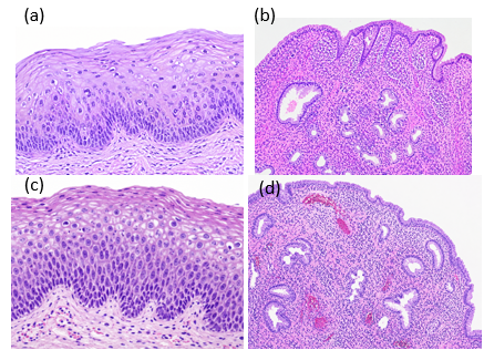
Figure 2: Representative histopathological findings of cervical lesions identified in the study. (a) CIN I; (b) chronic cervicitis; (c) CIN II; and (d) endocervical polyp. H&E staining
Overall, the findings demonstrate a broad histopathological spectrum among women undergoing cervical biopsy, ranging from inflammatory and benign lesions to premalignant and malignant conditions. Chronic cervicitis was the most frequently identified diagnosis, while cervical intraepithelial neoplasia constituted a substantial proportion of the specimens. The presence of both premalignant and malignant lesions highlights the clinical importance of timely biopsy and histopathological assessment. These findings provide the basis for interpreting the observed diagnostic patterns and their clinical relevance.
A total of 186 cervical biopsies were analysed in women aged 43.8 ± 11.6 years. The most common diagnosis was chronic cervicitis (29.6%), followed by premalignant lesions (45.2%). Malignant lesions (17.7%) were mostly squamous cell carcinoma (SCC), and were found to increase significantly after menopause. The age of 43.8 years was similar to that reported by Kaseka et al who reported a mean age of 41 years, cervicitis as the commonest non-malignant lesion, and SCC in 15.6% of all cervical biopsies 13. Similarly, Kerthi and Chander reported the highest incidence of cervical pathology at 41-50 years, chronic cervicitis being the most common non-neoplastic disease and SCC being the most common malignancy 14.
The burden of CIN was high, with CIN I, CIN II, and CIN III being 19.9%, 15.1% and 10.2% of biopsies, respectively. The overall incidence of premalignant lesions of 45.2% was significantly higher than 15% reported by Deepika et al. in 2026 15. The decreasing prevalence of CIN I to CIN III is consistent with the natural history of cervical neoplasia, as lower-grade lesions occur more frequently and have a greater chance of resolving. In the meta-analysis conducted by Loopik et al. in 2021, significant regression of CIN1 and CIN2 was observed, and as the grade of the lesion increased, its persistence also increased 16. In more recent pathology studies, there has also been a consistent increase in the expression of p16, Ki-67, and other markers of proliferation in CIN and invasive SCC 17.
There was a clinically significant association with age and presentation. The age distribution of pre-cancerous lesions was skewed towards women aged 30-49, while malignant lesions were more common the older the woman, and 45% of women aged ≥60 years had malignant lesions. Similarly, Sakai et al. showed age-related differences in the detection of CIN2+ by cytology and high-risk HPV testing 18. However, these findings are descriptive and do not establish an independent effect of age on histopathological severity. The most common symptom of the biopsy was abnormal vaginal bleeding (31.2%) followed by postcoital bleeding (22.0%). Rasool et al. reported that postcoital bleeding was associated with abnormal cervical cytology and emphasized appropriate cervical evaluation in women presenting with this symptom 19. The present study showed that abnormal Pap smears were associated with a high proportion of CIN I–III (76.4%). However, this descriptive finding does not by itself establish the diagnostic accuracy or effectiveness of cytology as a triage tool. Consistent with recent cytohistological correlation data, which noted a lack of sensitivity of cytological examination to detect glandular lesions, some glandular lesions might be missed by cytological examination and require tissue examination 20.
There was a statistically significant association between menopausal status and the grouped histopathological category, as the malignant group consisted of 37.1% of postmenopausal women and 8.1% of premenopausal women (p<0.001). This finding is significant because after menopause, the changes of epithelial atrophy and retraction of the transformation zone may make cytologic and/or colposcopy diagnosis more challenging 21. An older age was associated with pathological upgrading in women with CIN2/3, especially in those who had been postmenopausal longer and who were infected with HPV16/18, reported Jia et al. 22. The high-risk HPV and related clinical factors are also recently found to be predictors of high-grade lesions in postmenopausal HPV-positive women 23. These data justify a careful tissue diagnosis and a risk-based follow-up in older women with persistent cervical abnormalities.
The study is retrospective, single-center, and had a modest sample size, and relied on routine records, which can cause missing data and selection bias. There were no standardized additional biomarkers used with routine H&E histology, although recent evidence suggests the use of p16/Ki-67 and optimized biopsy strategies in certain diagnostically challenging cases 24. Multicenter prospective studies with HPV genotyping, cytology, colposcopy, standardized histopathological review and follow-up should be conducted to predict the persistence, progression and occurrence of invasive cancer.
In this retrospective biopsy series, cervical intraepithelial lesions formed the largest histopathological category, while chronic cervicitis was the most common individual diagnosis. Invasive malignancy occurred in 17.7% of specimens, with squamous cell carcinoma predominating. Malignancy was more frequent among postmenopausal women, although this unadjusted association may reflect age. Appropriate histopathological examination remains important for identifying high-grade and invasive disease. Future multicenter prospective studies should incorporate HPV testing, screening history, standardized cytology and colposcopy, and longitudinal follow-up.

Journal of Biomolecules, Pathogenesis and Therapeutics, J Biomol Pathog Ther. 2026;2(2), p69-71 (jbptjournal.org) © 2026 Authors. This work is published by Multidisciplinary Scholarly Advancement and Research MSAR Institute. The full terms of Journal Publishing policy is available at https://jbptjournal.org/index.php/jbpt/-publishing-license and incorporate the Creative Commons Attribution – Non Commercial (CC BY, NC 4.0) License https://creativecommons.org/licenses/by-nc/4.0/. By accessing the work you hereby accept the Terms. Non-commercial uses of the work are permitted without any further permission, provided the work is properly attributed. Publisher’s Note: MSAR Institute remains neutral with regard to jurisdictional claims in published maps and institutional affiliations, and assumes no liability for the scientific accuracy or clinical efficacy of the content herein, as they rest entirely with the authors.