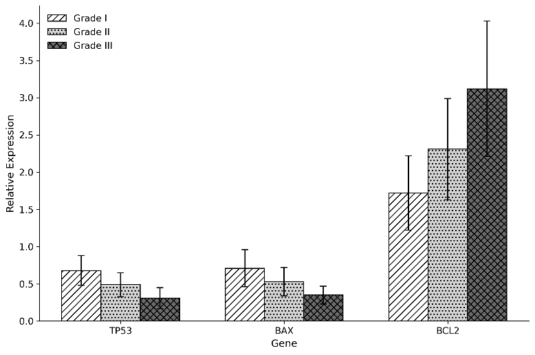
Figure 1. Relative expression of TP53, BAX, and BCL2 according to histological tumor grade.
Zainab Rehman1 | Muazzam Kashif2 | Qayyum Ahmed2 | Mavara Iqbal3*
1Department of Pathology, Chughtai Lab, Lahore, Pakistan | 2Department of Pathology, Superior University, Lahore, Pakistan
3Institute of Microbiology, UVAS, Lahore, Pakistan
*Correspondence: Mavara Iqbal ([email protected])
Received: 08 August, 2026; Revised: 05 September, 2026; Accepted: 11 September, 2026; Published: 20 September, 2026
Background: Disruption of genes that play a role in the regulation of apoptosis, both DNA-damage responses and mitochondrial cell death, can influence breast carcinoma progression. The objective of this study was to compare the expression of TP53, BAX, and BCL2 between histologically confirmed breast carcinoma and non-malignant tissues and to check the clinicopathological associations. Methods: A case-control analysis was performed, comprising 90 tissue samples, with breast carcinoma (60) and non-malignant (30). Total RNA was extracted and converted to complementary DNA (cDNA). The mRNA expression of TP53, BAX, and BCL2 was quantified by quantitative real-time PCR, normalized to GAPDH, and calculated using the 2^-ΔΔCt method. Growth patterns were assessed based on tumour grade, tumour size, and lymph-node involvement. Results: Carcinoma tissues showed lower TP53 (0.46 ± 0.18) and BAX (0.52 ± 0.21) and higher BCL2 (2.38 ± 0.74) expression than controls. TP53 and BAX decreased, whereas BCL2 increased, from Grade I to III tumors. The BAX/BCL2 ratio declined from 1.02 ± 0.18 in controls to 0.11 ± 0.05 in Grade III tumors. Gene expression was also associated with tumor size and lymph node involvement. Conclusions: Breast carcinoma showed downregulation of TP53 and BAX and upregulation of BCL2, with a progressive decline in the BAX/BCL2 ratio across tumor grades. These findings indicated altered apoptotic regulation associated with clinicopathological features.
Keywords: Apoptosis; bcl-2-Associated X Protein; Breast Neoplasms; Genes, bcl-2; Genes, p53; Real-Time Polymerase Chain Reaction
Breast cancer is one of the leading causes of cancer-related mortality and morbidity worldwide and, based on the most recent estimates of GLOBOCAN, accounts for about 11.8% of new cancer cases and 7.1% of cancer deaths in the world in 2024 1. It is a clinically diverse disease, depending on the histological type of the disease, the molecular subtypes, the genomic abnormalities, and the tumor microenvironment 2. In addition to continued proliferation and chromosomal instability, resistance to programmed cell death facilitates tumor progression, metastasis, and treatment failures 3.
TP53 and the BCL2 protein family are important regulators of the intrinsic apoptotic pathway 4. After recognizing damage to the DNA, functional p53 can trigger cell-cycle arrest, DNA repair, senescence, or apoptosis, in part by activating pro-apoptotic mediators, including BAX 5. TP53 mutations are present in approximately 1/3 of all breast cancers and are more prevalent in high-grade tumors, HER2-enriched tumors, and triple-negative breast cancers, which are aggressive and resistant to therapy 6. The BAX protein increases the permeability of the outer mitochondrial membrane and the release of cytochrome-c, while BCL2 inhibits the release of pro-apoptotic proteins and also helps to maintain the survival of the cells 7. Thus, the BAX/BCL2 ratio might be a better indicator of the apoptotic susceptibility than either of the markers separately 8. BCL2 expression in some cohorts can be associated with favorable hormone-receptor-positive disease, but its biological and prognostic significance is found to be dependent on the disease subtype and can be interpreted along with TP53, BAX and clinicopathological variables 9.
In parallel, studies that compare all four genes (TP53, BAX, BCL2 and the ratio of BAX to BCL2) at the tissue level in breast carcinoma, using quantitative methods at the transcript level, are limited. Integrated profiling could help elucidate the link between apoptotic dysregulation, tumour grade, size and lymph-node involvement 10. The findings may provide molecular insights into apoptotic dysregulation in breast carcinoma and help identify gene-expression patterns associated with clinicopathological characteristics.
The aim of this study was to quantify the expression of the genes TP53, BAX and BCL2 in breast carcinoma and healthy breast tissues by RT-qPCR. It was also designed to establish the apoptotic balance by measuring the BAX/BCL2 expression ratio. It also explored correlations of gene-expression profiles and selected clinicopathological features.
The study design and the collection of samples: A molecular case-control study was planned to investigate the expression profile of breast carcinoma tissues involving the genes related to apoptosis. These included 90 tissue samples, including 60 breast cancer samples and 30 healthy breast tissue samples from patients being operated on for breast cancer at authors' affiliated hospital. Sample size was calculated using OpenEpi version 3.0.1 (Emory University, Atlanta, GA, USA; RRID:SCR_021913) for an unmatched case-control study, assuming a two-sided confidence level of 95%, statistical power of 80%, and a case-to-control ratio of 2:1. Based on an expected exposure/proportion of 50% among controls and 75% among cases, the calculated minimum sample size was approximately 87 participants. To account for potential sample loss or inadequate RNA quality, 90 tissue samples were included 11. Before the molecular diagnosis of breast cancer cases, histopathological routine diagnosis was carried out.
Histopathological Confirmation: Routine histopathologic techniques were used to analyze the tumor samples. Tissues were fixed in 10% neutral buffered formalin, processed in the routine manner, embedded in paraffin, sectioned, and then stained with hematoxylin and eosin. The pathological diagnosis, the type of tumour and the tumour grade were noted based on standard pathological criteria. Data on the clinicopathological parameters, including tumour size, histological grade, lymph node status and receptor profile, were obtained from the pathology record.
Extraction of RNA and Quality Control of RNA: Fresh tissue specimens were immediately frozen in liquid nitrogen/placed in an RNA stabilization solution and stored at −80°C until RNA extraction. Total RNA was extracted from formalin-fixed paraffin-embedded (FFPE) tissue sections using the RNeasy FFPE Kit (QIAGEN, Germany) according to the manufacturer's instructions. The RNA concentration and purity were determined by spectrophotometric measurements. Samples of acceptable-quality RNA were selected for further analysis.
cDNA synthesis and quantitative real-time PCR: The extracted RNA was reverse transcribed using a cDNA synthesis kit (Merck) following the manufacturer's instructions. Quantitative real-time PCR was used to measure the gene expression of TP53, BAX, and BCL2. The endogenous reference gene for normalization was GAPDH. Gene-specific primers were used for amplification in the optimized cycling conditions, as presented in Table I. The comparative threshold cycle method (2-ΔΔCt) was used to calculate relative levels of gene expression. The expression level of tissues from breast cancer and control tissues was compared.
| Gene | Forward primer (5′–3′) | Reverse primer (5′–3′) |
|---|---|---|
| BAX | AGCTGCAGAGGATGATTGCC | CCCCAGTTGAAGTTGCCGTC |
| BCL2 | TGTGTGTGGAGAGCGTCAAC | CTACCCAGCCTCCGTTATCC |
| TP53 | GAGCTGAATGAGGCCTTGGA | CTGAGTCAGGCCCTTCTGTCTT |
Statistical Analysis: The data were analyzed using SPSS Statistics, Version 25.0 (IBM Corp., Armonk, NY, USA). The continuous variable data were presented as mean ± S.D., and the categorical variable data were presented as frequency and percent. Statistical comparisons were used to determine the differences in the expression of each group. A correlation was made between the expression levels of the genes and the different clinicopathological parameters. A p-value < 0.05 was considered statistically significant. The study protocol is in line with the institutional ethical guidelines. Molecular data analysis was performed without any personal identifiers of patients, in order to ensure patient confidentiality.
A total of 90 tissues (60 breast carcinomas and 30 non-malignant breast tissue controls) were tested. The relative gene expression analysis was done by the 2^-ΔΔCt method, using GAPDH as an endogenous control. Analysis demonstrated differential expression of the genes related to apoptosis in the course of malignant tissue versus control tissue. Breast carcinoma samples indicated lower expression levels for tumor suppressor and pro-apoptotic genes, and elevated expression levels for the BCL2 gene as compared to the control samples. The clinical characteristics of patients with breast cancer used for molecular analysis are shown in Table II. Most cases were in women of middle age (40s), and the most common histological type was invasive ductal carcinoma. Tumor grade distribution showed a greater representation of intermediate-grade tumors.
| Characteristics | Frequency (n) | Percentage (%) |
|---|---|---|
| Age group | ||
| <40 years | 18 | 30.0 |
| 40–60 years | 32 | 53.3 |
| >60 years | 10 | 16.7 |
| Histological type | ||
| Invasive ductal carcinoma | 49 | 81.7 |
| Invasive lobular carcinoma | 7 | 11.7 |
| Other types | 4 | 6.6 |
| Histological grade | ||
| Grade I | 12 | 20.0 |
| Grade II | 31 | 51.7 |
| Grade III | 17 | 28.3 |
| Lymph node involvement | ||
| Present | 26 | 43.3 |
| Absent | 34 | 56.7 |
The extent of the variation in the expression of these genes for apoptosis is indicated in Table III when comparing malignant tissue with non-malignant tissue. TP53 and BAX were also seen to be down-regulated, and BCL2 was up-regulated in breast cancer samples, indicating over-suppression of pro-apoptotic genes and over-activation of survival genes in tumor samples.
| Gene | Control Tissue (Relative Expression) | Breast Cancer Tissue (Relative Expression) | Expression Pattern |
|---|---|---|---|
| TP53 | 1.00 ± 0.12 | 0.46 ± 0.18 | Downregulated |
| BAX | 1.00 ± 0.15 | 0.52 ± 0.21 | Downregulated |
| BCL2 | 1.00 ± 0.14 | 2.38 ± 0.74 | Upregulated |
The expression patterns of the apoptosis-related genes according to tumor grade are shown in Table IV. The expression of TP53 and BAX was found to be decreasing with the increasing grade of the tumour, while that of BCL2 was increasing. These findings indicate an altered apoptotic gene-expression pattern in higher-grade tumors.
| Gene | Grade I | Grade II | Grade III | Trend |
|---|---|---|---|---|
| TP53 | 0.68 ± 0.20 | 0.49 ± 0.16 | 0.31 ± 0.14 | Decreased expression with higher grade |
| BAX | 0.71 ± 0.25 | 0.53 ± 0.19 | 0.35 ± 0.12 | Reduced pro-apoptotic activity |
| BCL2 | 1.72 ± 0.50 | 2.31 ± 0.68 | 3.12 ± 0.91 | Increased anti-apoptotic activity |
Figure 1 illustrates the relative expression of TP53, BAX, and BCL2 according to histological tumor grade. TP53 expression decreased progressively from Grade I to Grade III tumors, while BAX expression showed a similar downward pattern. In contrast, BCL2 expression increased with increasing tumor grade. The figure therefore demonstrates distinct expression patterns of the three apoptosis-related genes across different histological grades.
Figure 1. Relative expression of TP53, BAX, and BCL2 according to histological tumor grade.
The associations between gene-expression patterns and clinicopathological features are shown in Table V. Associations between the changes in expression of apoptosis-related genes and the size of the tumor, histological grade, and lymph node involvement suggest differential involvement of pro-apoptotic and anti-apoptotic pathways in breast carcinoma progression.
| Variable | TP53 Expression | BAX Expression | BCL2 Expression |
|---|---|---|---|
| Tumor size >2 cm | Lower expression | Lower expression | Higher expression |
| High histological grade | Reduced | Reduced | Increased |
| Lymph-node involvement | Reduced | Reduced | Increased |
A balance between pro-apoptotic and anti-apoptotic pathways is emphasized in Table VI. The ratio of BAX/BCL2 decreased in the malignant tissues compared with control tissue and became further decreased with an increase in the grade of the cancer. The progressive decline in the BAX/BCL2 ratio indicates a relative shift toward anti-apoptotic signaling in higher-grade tumors.
| Group | BAX/BCL2 Ratio | P- value |
|---|---|---|
| Control tissues | 1.02 ± 0.18 | <0.001 |
| Breast cancer tissues | 0.22 ± 0.09 | 0.001 |
| Grade I tumors | 0.41 ± 0.13 | <0.001 |
| Grade II tumors | 0.24 ± 0.08 | <0.001 |
| Grade III tumors | 0.11 ± 0.05 | <0.001 |
The relationships between the gene expression levels and pathological parameters are compared, as shown in Table VII. TP53 was inversely associated with aggressive tumor features, BAX was inversely associated with aggressive tumor features, and BCL2 was positively associated with aggressive tumor features, indicating inverse associations of TP53 and BAX expression with aggressive tumor features and a positive association of BCL2 expression with these features.
| Parameter | TP53 (ρ) | BAX(ρ) | BCL2(ρ) |
|---|---|---|---|
| Tumor grade | -0.61 | -0.56 | +0.64 |
| Tumor size | -0.42 | -0.39 | +0.45 |
| Lymph node involvement | -0.51 | -0.48 | +0.55 |
Overall, the expression profile demonstrated a consistent shift in the balance between pro-apoptotic and anti-apoptotic signaling with increasing tumor aggressiveness. Reduced TP53 and BAX expression, together with increased BCL2 expression, was associated with larger tumors, higher histological grade, and lymph-node involvement. The progressive reduction in the BAX/BCL2 ratio further supports an apoptosis-resistant molecular pattern in breast carcinoma. These findings provide the basis for interpreting the potential clinical relevance of these apoptosis-related genes.
This study shows altered expression of apoptosis-related genes in histologically proven breast carcinomas. The expression levels for TP53 and BAX were found to be 0.46-fold and 0.52-fold, respectively, of the non-malignant tissue, while BCL2 was 2.38-fold the expression level in non-malignant tissue. In the control, the ratio of BAX/BCL2 was 1.02, while it was 0.22 in carcinomas and was reduced gradually according to the grade of the tumor (from grade I to grade III). This is similar to the mitochondrial apoptosis suppression model, where impaired p53 signaling and BAX activity enable anti-apoptotic BCL2 proteins to maintain mitochondrial integrity 12. A 2024 long-term follow-up study similarly demonstrated that TP53 mutations were associated with significantly poorer recurrence-free and overall survival in patients with breast cancer 13. Studies also found that abnormal p53 immunophenotypes closely predicted TP53 mutation, and were related to high-grade triple-negative breast carcinomas 14.
Decreased transcript levels may lead to less activation of apoptotic targets like BAX, allowing genetically damaged cells to live and gain further oncogenic mutations. This interpretation is consistent with recent clinical evidence showing that a null p53 immunophenotype is associated with poorer disease-free and distant recurrence-free survival in patients with invasive breast cancer 15. Recent studies also suggest that TP53 dysfunction contributes to invasion, metabolic adaptation, immune escape, and resistance to systemic therapy 16,17. However, low TP53 mRNA is not synonymous with mutation, as there are truncating mutations, copy-number loss, epigenetic repression, and RNA degradation that can lead to low transcripts 18. The simultaneous downregulation of TP53 and BAX, therefore, facilitates functional pathway suppression and should be confirmed at the protein level and sequenced 19.
The most prominent change in the apoptotic balance was the combination of BAX downregulation and BCL2 upregulation. BAX is able to form pores in the mitochondria to allow the release of cytochrome-c, while BCL2 inhibits BAX and other pro-apoptotic proteins 20. Therefore, the decreasing BAX/BCL2 ratio would be indicative that high-grade tumors might need a more potent stimulus to trigger mitochondrial apoptosis. Experimental research in breast cancer cells also demonstrated that an increased BAX/BCL2 ratio, accompanied by increased caspase-7 and caspase-9 expression, promoted caspase-dependent apoptosis 21. Sofi et al. found that BCL2 overexpression is linked to survival pathways and chemoresistance, while Hassan et al. found both favorable and unfavorable associations in hormone receptor-positive and HER2-enriched tumors, respectively 22,23.
The correlations with grade were greater than those with tumour size and nodal involvement, indicating that the apoptotic dysregulation is more likely to be related to biological dedifferentiation. Evaluating TP53 and BAX, BCL2 and their ratio simultaneously will, therefore, be more informative than evaluating each of them individually. In a 2024 study that included 5,186 patients, Arg72Pro and Pro72Pro genotype were found to be associated with stable disease, but not pathological complete response (pCR) 24. Combined p53 and BCL2 immunophenotypes were also associated with prognosis in an age- and subtype-dependent manner in postmenopausal TNBC 25. Therefore, the TP53, BAX and BCL2 panel is still in its exploratory stage, and stage, grade, receptor subtype and lymph-node status are still known prognostic factors 26.
Some of the limitations of this study are the relatively small sample size, the case-control design, the lack of survival and treatment-response data, absence of protein-level validation, absence of TP53 mutation analysis and the use of bulk-tissue RT-qPCR, with GAPDH as the only endogenous control. To improve future multicenter studies, it is recommended to employ larger cohorts of patients stratified by subtype, paired controls, several validated reference genes, and combined RT-qPCR, immunohistochemistry, digital PCR, and sequencing. Integration of RT-qPCR with protein-level validation, multiple reference genes, TP53 sequencing, and functional assays of apoptosis would provide stronger evidence regarding the biological significance of these expression patterns. It will be important to find out whether this profile is independently associated with the likelihood of recurrence, therapeutic response, survival, or susceptibility to cell death-inducing drugs in longitudinal and functional analyses.
This study demonstrated reduced TP53 and BAX mRNA expression and increased BCL2 expression in breast carcinoma, with a lower BAX/BCL2 ratio. These patterns were associated with higher tumour grade, size, and lymph-node involvement, suggesting altered apoptotic regulation. However, as only mRNA expression was assessed, the findings remain exploratory and do not establish functional apoptosis resistance or tumour aggressiveness. Larger multicenter studies incorporating mutation, protein, functional, treatment-response, and survival analyses are warranted to determine the clinical and prognostic significance of this gene-expression profile.

Journal of Biomolecules, Pathogenesis and Therapeutics, J Biomol Pathog Ther. 2026;2(2), p69-71 (jbptjournal.org) © 2026 Authors. This work is published by Multidisciplinary Scholarly Advancement and Research MSAR Institute. The full terms of Journal Publishing policy is available at https://jbptjournal.org/index.php/jbpt/-publishing-license and incorporate the Creative Commons Attribution – Non Commercial (CC BY, NC 4.0) License https://creativecommons.org/licenses/by-nc/4.0/. By accessing the work you hereby accept the Terms. Non-commercial uses of the work are permitted without any further permission, provided the work is properly attributed. Publisher’s Note: MSAR Institute remains neutral with regard to jurisdictional claims in published maps and institutional affiliations, and assumes no liability for the scientific accuracy or clinical efficacy of the content herein, as they rest entirely with the authors.