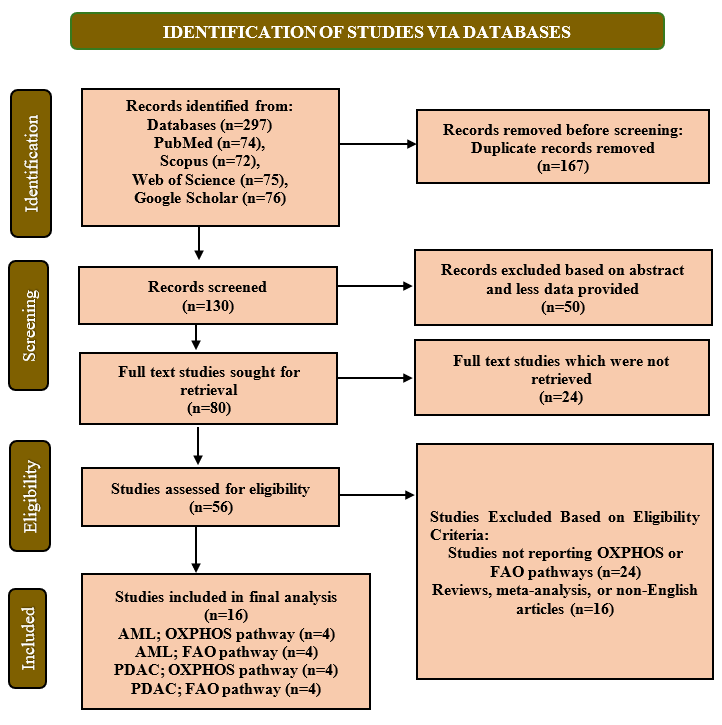
Figure 1: PRISMA Flow Diagram for Study Selection. The flowchart was designed according to the PRISMA guidelines 2020, showing study identification, screening, eligibility assessment, and final selection in the systematic review (n= 16)
Mahnoor Khan1* | Adil Kaleem1 | Abu Bakar Rehman1
1Department of Allied Health Sciences, Superior University, Lahore, Pakistan
*Correspondence: Mahnoor Khan ([email protected])
Received: 21 May, 2026; Revised: 01 September, 2026; Accepted: 13 September, 2026; Published: 20 September, 2026
Background: Mitochondrial metabolic reprogramming by Oxidative phosphorylation (OXPHOS) and Fatty acid oxidation (FAO) has become survival and therapy-resistance driver in acute myeloid leukemia (AML) and pancreatic ductal adenocarcinoma (PDAC). This review aimed to evaluate the role of OXPHOS and FAO pathways in cancers and potential as therapeutic targets. Methods: The research was conducted according to PRISMA 2020 guidelines. PubMed, Scopus, Web of Science, and Google Scholar databases were searched from January 2013 to December 2025. Articles with data on effect of pharmacological or genetic inhibition of OXPHOS and FAO in AML and PDAC were considered. Reviews, case reports, and non-English articles were excluded. The SYRCLE and OHAT tools were used to determine the risk of bias, and the certainty of evidence was determined using GRADE. MetaAnalysisOnline Tool was used for statistical analysis. Results: Sixteen studies met inclusion criteria. Studies included in AML assessed OXPHOS (SMD = -0.76, 95% CI: -3.18 to -1.67, p= 0.05) and FAO (SMD = -0.19, 95% CI: -7.31 to -6.93, p> 0.05) showed no significant differences and high heterogeneity (I2 > 94%). In PDAC, four studies provided data on OXPHOS (SMD = -5.27, 95% CI: -15.59 to -5.05, p .05) and four on FAO (SMD = -215.42, 95% CI: -616.27 to -185.44, p>0.05) and high heterogeneity (I² > 95%). The overall risk of bias was low to moderate, and certainty of evidence was moderate. Conclusions: In cancer cells, AML, and PDAC, OXPHOS and FAO were parallel dependent pathways, making mitochondrial metabolism a promising target therapy.
Keywords: Leukemia, Myeloid, Acute; Metabolic Reprogramming; Molecular Targeted Therapy; Oxidative Phosphorylation; Pancreatic Neoplasms
Cancer cells undergo extensive metabolic reprogramming to facilitate unregulated growth, survival during metabolic stress, and resistance to therapy. Even though enhanced glycolysis had been regarded as a feature of cancer metabolism, mitochondrial metabolism was currently acknowledged as a primary determinant of tumor progression and resistance to therapy 1,2. This was particularly evident in cancers that rely on mitochondrial respiration to meet energy, redox, and biosynthetic demands, often using non-glycolytic substrates such as fatty acids. Mitochondrial metabolism is especially important in aggressive malignancies, including acute myeloid leukemia (AML) and pancreatic ductal adenocarcinoma (PDAC) 3.
AML and PDAC are two types of aggressive cancers with poor prognosis, where mitochondrial dependencies play important roles. Leukemic stem cells (LSCs) in AML, particularly in relapsed or chemo-resistant subpopulations, were observed to be more dependent on OXPHOS to live and survive 4. FAO often sustained this dependency and provided acetyl-CoA to the tricarboxylic acid (TCA) cycle and fueled the electron transport chain (ETC), leading to therapy resistance and LSC persistence 5. Similarly, in PDAC, which was considered one of the most fatal solid tumors, mitochondrial metabolism became a major vulnerability. Cancer stem cells (CSCs) were subsets of PDAC cells that had active OXPHOS and required FAO for the production of ATP in nutrient-deficient or stressful conditions 6-8. FAO inhibition disrupted OXPHOS, caused energy crises, and reduced tumorigenicity and chemoresistance in PDAC models. This metabolic plasticity enabled PDAC cells to adapt to hostile tumor microenvironments, highlighting OXPHOS and FAO as potential therapeutic targets 9. Although interest in mitochondrial metabolic reprogramming in cancer is increasing, evidence on OXPHOS and FAO in AML and PDAC remains fragmented, and systematic comparison of shared metabolic dependencies, regulatory mechanisms, and therapeutic strategies remains limited. Targeting OXPHOS components, FAO enzymes such as CPT1, or upstream regulators may help overcome resistance and improve treatment outcomes 10.
This systematic review and meta-analysis aimed to synthesize current evidence on the dual mitochondrial dependencies involving OXPHOS and FAO in AML and PDAC. It studied the molecular mechanisms underlying these dependencies, their role in pathogenesis and resistance, and new approaches to reprogramming these pathways therapeutically to increase anti-cancer efficacy.
Study Design and Reporting Standards: This systematic review and meta-analysis was conducted in accordance with the PRISMA 2020 guidelines 11.
Inclusion and Exclusion Criteria: Studies were thoroughly evaluated in terms of inclusion and exclusion criteria by two independent reviewers. Original research articles, including experimental preclinical studies and clinical translational studies that investigated quantitative measurements of mitochondrial metabolic activity related to OXPHOS or FAO in AML and PDAC were included. Reviews, meta-analyses, editorials, case reports, conference abstracts, non-English publications, and studies with no relevant outcome data or insufficient quantitative findings were excluded.
Data Sources: Four databases, including PubMed, Scopus, Web of Science, and Google Scholar, were systematically searched to identify relevant studies published from January 2013 to December 2025.
Search Strategy: The search strategy used a combination of MeSH terms and free-text keywords related to “Acute Myeloid Leukemia”, “Pancreatic cancer”, “Oxidative phosphorylation”, “Fatty acid oxidation”, and “mitochondrial reprogramming”. Boolean operators AND and OR were combined with these search terms to refine the search and reference lists of included studies were checked manually to identify other relevant articles.
Study Selection: Two independent reviewers screened each study's title and abstract until the appropriate inclusion requirement was satisfied. Full-text articles were subsequently obtained for potentially eligible studies and evaluated independently. The reviewers used expert consultation or group consensus to resolve their disagreements over selection.
Data Extraction: Data extraction was performed using a standardized form comprising the following variables: first author, publication year, study design ad cancer type, sample size and study characteristics, mitochondrial reprogramming pathway investigated, key findings, and therapeutic implications. Quantitative findings, including Mean and Standard Deviation (SD), were extracted from each study. Studies reporting zero SD were adjusted using a continuity correction (SD = 0.0001) to allow inclusion in inverse-variance meta-analysis.
Outcome Measures: The primary outcome of this review and meta-analysis was to evaluate the role of mitochondrial OXPHOS and FAO pathways in metabolic reprogramming, tumor progression, and targeting these pathways for therapeutic implications of AML and PDAC. The studies were categorized based on cancer type and mitochondrial metabolic pathway investigated. The secondary outcomes included assessment of pathway-specific effects on ATP production, mitochondrial respiration, cancer cell viability, tumour growth, therapy resistance, and therapeutic response after OXPHOS or FAO inhibition. Because the included studies used different experimental designs, meta-analysis was performed only when studies were sufficiently comparable in cancer type, pathway, intervention/comparator, outcome, and measurement method. Where appropriate, analyses were stratified by study design, cancer type, and pathway to account for methodological heterogeneity.
Quality Assessment: Risk of bias (ROB) of included studies was assessed through the SYRCLE Risk of Bias Tool for animal experimental studies and the OHAT risk of bias tool for in vitro mechanistic studies 12,13. Risk of bias ratings were visualized using traffic light plots. The overall certainty of evidence was assessed using the GRADE approach.
Data Synthesis: Statistical analysis, including generation of forest plots and funnel plots, was performed using MetaAnalysisOnline.com, Version 1 (A5 Genetics Ltd., Hungary; web-based statistical tool for meta-analysis) 14. From each study, mean and SD were extracted, and pooled effect sizes, specifically Standardized Mean Difference (SMD) with 95% confidence intervals (CI), were calculated to compare metabolic markers between experimental (treated/knockdown) and control groups. Heterogeneity was assessed using the I² statistic, with values interpreted as low (≤50%), moderate (50–75%), or high (>75%). Subgroup analysis was performed to find possible sources of variation according to study design, disease subtype, and clinical characteristics. For outcomes where statistical pooling was precluded due to extreme heterogeneity, a narrative synthesis was provided. Sensitivity analysis was conducted by sequentially omitting individual studies to evaluate the robustness of the results. Fifteen studies were included for quantitative analysis. Studies reporting zero SD were adjusted using a continuity correction (SD = 0.0001) to allow inclusion in inverse-variance meta-analysis.
Figure 1: PRISMA Flow Diagram for Study Selection. The flowchart was designed according to the PRISMA guidelines 2020, showing study identification, screening, eligibility assessment, and final selection in the systematic review (n= 16)
Following an extensive search across four electronic databases and supplementary sources, a total of 297 research articles were initially identified. After removing 167 duplicates, 130 records remained. Title and abstract screening excluded 50 studies, leaving 80 articles for full-text review. Of these, 24 were inaccessible, and a further 56 were excluded due to lack of stratified data, inappropriate study design (e.g., reviews, case reports), or non-English language. Ultimately, sixteen studies met the inclusion criteria and were included in this schematic review examining OXPHOS and FAO pathways in AML and PDAC. The PRISMA 2020 flow diagram presented in Figure 1 illustrated the study selection process.
Study characteristics of included studies, such as first author and publication year, study design, cancer type, sample size, targeted mitochondrial metabolic pathway (OXPHOS or FAO), key findings, and therapeutic implications of each study were summarized in Table I. The selected articles assessed mitochondrial metabolic requirements and therapeutic susceptibilities of the OXPHOS pathways and fatty acid oxidation of acute myeloid leukemia and pancreatic ductal adenocarcinomas.
Table I: Characteristics of Included Studies with Key Outcomes, Study Design, Sample Size, Quantitative Data, and Key Findings
| Author & Year | Study design (Cancer type) | Sample Size (Study Characteristics/ Scheme of Groups) | Mitochondrial Reprogramming Pathway | Outcomes/ Key findings | Therapeutic implications/ Interventions | Quantitative findings |
|---|---|---|---|---|---|---|
| Pollyea et al., 2018 15 | Clinical translational study (AML) | n =33 (Primary human AML cells, human AML patients) | OXPHOS pathway | By blocking the metabolism of amino acids and lowering OXPHOS, venetoclax and azacitidine dramatically decreased the viability of LSCs in AML | Targeting OXPHOS-dependent LSC with combined venetoclax–azacitidine therapy acts as major therapeutic target in AML | Mean ± SD (pmol/min) Control: 1 ± 0.0001 Treatment: 0.47 ± 0.80 |
| Lagadinou et al., 2013 16 | Experimental study (AML) | n=5 (Normal and leukemic human bone marrow samples), n=10/group (NSG mice) | OXPHOS pathway | By inhibiting OXPHOS, BCL-2 inhibition specifically targeted quiescent LSCs | OXPHOS-dependent LSCs were specifically eliminated by BCL-2 inhibitors, indicating that mitochondrial metabolism represents a selective vulnerability in AML | Mean ± SD (nM) Control: 89.2 ± 5.59 Treatment: 43.2 ± 12.75 |
| Yang et al., 2017 17 | Experimental study (AML) | n=14 (OCI-AML3 cells) | OXPHOS pathway | IACS-010759 inhibitor causes AMPK activation induced apoptosis of AML cells | OXPHOS inhibitor act as an important biomarker of anti-leukemic activity | Mean ± SD (nM) Control: 0.5 ± 0.37 Treatment: 8.6 ± 15.71 |
| Banellaet al., 2022 18 | Experimental study (AML) | n=17 (AML bone marrow samples), (OCI-AML3, OCI-AML2 and U937-AETO cell lines) | OXPHOS pathway | Ascorbate + buformin impaired mitochondrial respiration and reduced AML viability | Combination therapy targets OXPHOS metabolism in AML | Mean ± SD (pmol/min) Control: 8 ±1 Treatment: 23 ± 16 |
| Yang et al., 2025 19 | Experimental study (AML) | n=5 (AML2, K562, HL-60, KG1a cell lines) | FAO Pathway | In AML cells, pharmacological inhibition of PPARδ by 6ME specifically reduced FAO, resulting in reduced ATP synthesis, mitochondrial stress, and death | A unique method for causing selective mortality in AML is pharmacological inhibition of PPARδ with 6ME | Mean ± SD (μM) Control: 1000 ± 0.0001 Treatment: 0.8748 ± 0.176 |
| Farge et al., 2017 20 | Experimental + Translational (AML) | n=21 (AML samples), (MOLM14 cells lines) | FAO Pathway | FAO inhibition in chemotherapy-resistant AML cells with Etomoxir combined with AraC reduced oxygen consumption and fatty acid oxidation rate | Combined targeting of OXPHOS and FAO metabolism may eliminate chemotherapy-resistant AML cells and reduce relapse risk. | Mean ± SD (μM) Control: 0.10 ± 0.0046 Treatment: 0.013 ± 0.0183 |
| Tcheng et al., 2022 21 | Experimental study (AML) | n=3 (Patient derived AML and normal MNC cells), (TEX and OCl-AML2cell lines) | FAO Pathway | LSC viability was lowered by AYNE's potent inhibition of FAO and reduction of mitochondrial energy generation | Natural FAO inhibitors such as AYNE may represent novel mitochondrial metabolic therapies targeting FAO-dependent LSCs | Mean ± SD (μM) Control: 1 ± 5 Treatment: 0.78 ± 4 |
| Tabe et al., 201822 | Experimental mechanistic study (AML) | (THP-1, MOLM13 and OCI-AML3 cell lines) | FAO Pathway | Avocatin B treatment significantly decreased the levels of FAO cycle and marked repression of OCR | FAO inhibition (etomoxir) sensitized AML cells to cytarabine (AraC); combination therapy enhanced apoptosis and reduced metabolic activity | Mean ± SD pmol/min) Control: 330 ± 50 Treatment: 74 ± 30 |
| Yu et al., 2019 23 | Experimental study (PDAC) | n=10 (mouse xenograft models), (MDA-PATC118 and HPNE cell lines) | OXPHOS pathway | Direct mitochondrial fusion by MFN2 expression reduced OCR basal respiration, and ATP production | Encouraging mitochondrial fusion takes advantage of PDAC's distinct mitochondrial biology to trigger mitophagy reducing tumor | Mean ± SD pmol/min) Control: 165 ± 10 Treatment: 68 ± 8 |
| Xue et al., 202224 | Experimental + translational study (PDAC) | n=5/group (C57BL/6 mice), (MIA PaCa-2 cells) | OXPHOS pathway | DX3–213B is a highly potent OXPHOS complex I inhibitor which impairs ATP generation, and blocks MIA PaCa-2 cell growth | Different compounds can be used for OXPHOS inhibition, which can be a safe and efficacious strategy to treat pancreatic cancer | Mean ± SD (nM) Control: 0.07 ± 0.01 Treatment: 11 ± 1.5 |
| Wang et al., 2020 25 | Experimental study (PDAC) | n=8/group (xenografts mouse models) | OXPHOS pathway | UQCRC1 knockdown reduced OXPHOS and tumor progression | UQCRC1 is a potential OXPHOS-targeted therapeutic biomarker in PDAC | Mean ± SD (nM) Control: 0.86 ± 0.07 Treatment: 0.51 ± 0.09 |
| Dash et al., 2024 26 | Experimental study (PDAC) | n=18 (BALB/cAJcl-nu/nu mice), (CFPAC-1 cell lines) | OXPHOS pathway | Mitochondrial metabolism inhibition via dCK inactivation reduced viability of resistant pancreatic cancer cells | Enetoclax and a mitochondrial complex I inhibitor are therapeutically efficacious for cancerous cells | Mean ± SD (nmol/L) Control: 2.9 ± 0.1 Treatment: 1.75 ± 0.02 |
| Woo et al., 2025 27 | Experimental study (PDAC) | n=52 (Slc25a20 knockout mice, Balb/c-nu/nu mice), n=164 (n = 27; normal tissues, n = 137; PDAC patient tissues) | FAO Pathway | Loss of SLC25A20 decreased the development of tumor, ATP production and compromised mitochondrial FAO | SLC25A20 inhibition targeting mitochondrial FAO pathway is a possible metabolic treatment approach against PDAC | Mean ± SD (nM) Control: 1 ± 0.0001 Treatment: 0.4 ± 0.001 |
| Sharma et al., 2024 28 | Experimental + translational study (PDAC) | n=3 (MIA PaCa-2 cells ), (PANC-1 and MIA PaCa-2, FC-1199, HPNE cell lines) | FAO Pathway | Compounds 6 (methyl ester) showed comparable antiproliferative effects against PANC-1 and MIA PaCa-2 cell lines | The lead compounds inhibit the proliferation of human pancreatic cancer cell lines, exhibiting a synergistic cytotoxic effect with the OXPHOS inhibitor phenformin | Mean ± SD (μM) Control: 100 ± 0.0001 Treatment: 80 ± 17.3 |
| Shinoda et al., 2025 29 | Experimental study (PDAC) | n=15 (AKO mice), n=6 (C57BL/6J mice), (KPC cells, murine pancreatic cancer cell lines, PANC-1, CAPAN-2, HT29, and GIST-T1 cell lines) | FAO pathway | FABP inhibition demonstrated reliance on FA metabolism by decreasing its usage and inhibiting pancreatic tumor growth and metastasis | Targeting fatty acid transport and FAO metabolism may represent a therapeutic strategy to inhibit pancreatic cancer progression and metastasis | Mean ± SD (μM) Control: 1 ± 0.02 Treatment: 0.5 ± 0.01 |
| Lee et al., 2020 30 | Experimental in vitro study (PDAC) | n=6/ group (Balb/c-nu mice), (PANC-1, MIA PaCa-2, BxPC-3 cell lines) | FAO Pathway | FAO inhibition via trimetazidine reduced ATP production and cell survival in PDAC cells | Targeting mitochondrial FAO metabolism may represent an effective metabolic therapeutic strategy in PDAC | Mean ± SD (μM) Control: 1 ± 0.0001 Treatment: 0.6 ± 1.46 |
| AML= Acute Myeloid Leukemia; PDAC= Pancreatic Ductal Adenocarcinoma; OXPHOS= Oxidative Phosphorylation; FAO= Fatty Acid Oxidation; LSC= Leukemic Stem Cell; SD= Standard Deviation; NSG mice= Non-Obese Diabetic Severe Combined Immunodeficiency Gamma mice; BCL-2= B-cell Lymphoma 2; AMPK= AMP-Activated Protein Kinase; ATP= Adenosine Triphosphate; PPARδ= Peroxisome Proliferator-Activated Receptor Delta; OCR= Oxygen Consumption Rate; AraC= Cytarabine (Arabinosyl Cytidine); MNC= Mononuclear Cells; TEX= TEX Leukemia Cell Line; MFN2= Mitofusin 2; NAD+/NADH= Nicotinamide Adenine Dinucleotide (oxidized/reduced forms); UQCRC1= Ubiquinol-Cytochrome c Reductase Core Protein 1; dCK= Deoxycytidine Kinase; SLC25A20= Solute Carrier Family 25 Member 20; FABP= Fatty Acid Binding Protein; AKO mice= Acyl-CoA Oxidase Knockout mice; KPC= KrasLSL-G12D/+; Trp53LSL-R172H/+; Pdx1-Cre mouse model, HTS= High-Throughput Screening, μM= Micromolar; nM= Nanomolar; mM= Millimolar; nmol/L= Nanomoles per Liter; nmole/mg= Nanomoles per Milligram; pmol/min= Picomoles per Minute. | ||||||
The included studies demonstrated that mitochondrial metabolic reprogramming through OXPHOS and FAO plays a crucial role in the survival and progression of both AML and PDAC. Several studies reported that inhibition of OXPHOS reduced mitochondrial respiration, ATP production, and cancer cell viability, while also inducing metabolic stress and apoptosis. In AML, targeting OXPHOS with agents such as venetoclax, azacitidine, and complex I inhibitors selectively impaired leukemic stem cell survival. Similarly, in PDAC models, inhibition of mitochondrial respiration or knockdown of mitochondrial proteins suppressed tumor growth and metabolic activity. In addition, multiple studies highlighted the role of FAO in maintaining tumor energy metabolism, where pharmacological or genetic inhibition of FAO reduced ATP production, mitochondrial function, and cancer cell proliferation. Overall, these findings indicate that targeting mitochondrial OXPHOS and FAO pathways may represent promising metabolic therapeutic strategies for AML and PDAC.
A total of fifteen studies were included in the meta-analysis. Tabe et al., 201822 was excluded from the meta-analysis as it only included cell lines, and there was no sample size mentioned in the study, so it was not possible to perform quantitative analysis on it. Studies reporting zero SD were identified during data extraction. To avoid disproportionate weighting and distortion of pooled estimates, these studies were excluded from quantitative pooling where a reliable variance estimate could not be obtained and were retained for qualitative synthesis.
There were four studies reporting OXPHOS pathway in AML that were analyzed with a total of 74 subjects in the Treatment cohort and 74 subjects in the Control cohort. Based on the analysis performed using a random-effects model with the inverse-variance method to compare the standardized mean difference (SMD), there was no statistical difference between the two cohorts (SMD = -0.76, 95% CI: -3.18 to - 1.67, p > 0.05). Significant heterogeneity was detected (p<0.01, I2 =94.1%), suggesting inconsistent effects across studies, as shown in Figure 2.
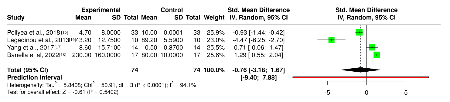
Figure 2: Forest plot comparing Mitochondrial Metabolic Activity between experimental treatment groups and controls in Acute Myeloid Leukemia (AML) models across various interventions targeting Oxidative Phosphorylation (OXPHOS) pathways in AML (n= 74)
Funnel plot for studies evaluating OXPHOS-targeting interventions in AML. The distribution of the study estimates was assessed to explore the possibility of publication bias. The plot did not indicate an obvious asymmetry suggestive of publication bias, as shown in Figure 3.
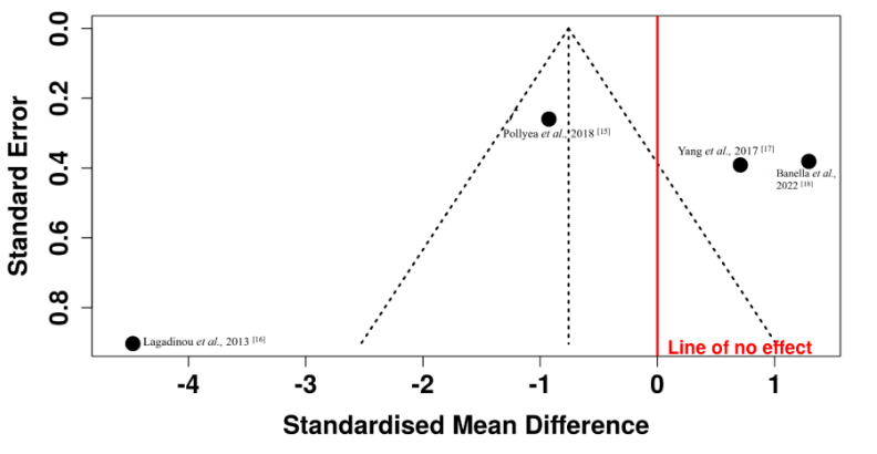
Figure 3: Funnel plot assessing publication bias for studies targeting Oxidative Phosphorylation (OXPHOS) pathway in Acute Myeloid Leukemia (AML) (n= 74)
Three studies focused on the FAO pathway in AML were analyzed, with a total of 29 subjects in the Treatment cohort and 29 subjects in the Control cohort. The analysis was performed as shown in Figure 4. There was no statistical difference between the two groups (SMD = -0.19, 95% CI: -7.31 to – 6.93, p > 0.05). Significant heterogeneity was detected (p<0.01, I2 = 96.4%), suggesting inconsistent effects across studies.
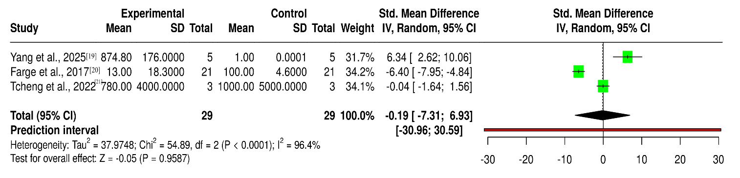
Figure 4: Forest plot comparing Mitochondrial Metabolic Activity between experimental treatment groups and controls in Acute Myeloid Leukemia (AML) models across various interventions targeting Fatty acid Oxidation (FAO) pathway in AML (n= 29)
Figure 5 presents the funnel plot for the studies assessing FAO-targeting interventions in AML. The individual study estimates are distributed around the pooled effect with varying levels of precision. Visual inspection of the funnel plot did not demonstrate an obvious asymmetry that would suggest substantial publication bias.
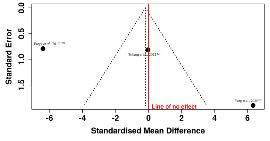
Figure 5: Funnel plot assessing publication bias for studies in Fatty acid Oxidation (FAO) pathway in Acute Myeloid Leukemia (AML) (n= 29)
For the OXPHOS pathway in PDAC, four studies were analyzed with a total of 41 subjects in the Treatment cohort and 41 subjects in the Control cohort. Based on the analysis performed, as shown in Figure 6, there was no statistical difference between the two cohorts (SMD = -5.27, 95% CI: -15.59 to -5.05, p > 0.05). Significant heterogeneity was detected (p<0.01, I2 =95%), suggesting inconsistent effects across the studies.
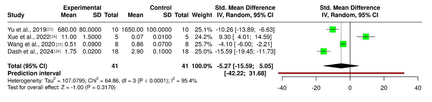
Figure 6: Forest plots comparing Mitochondrial Metabolic Activity between experimental treatment groups and controls in Pancreatic Ductal Adenocarcinoma (PDAC) models across various interventions targeting Oxidative Phosphorylation (OXPHOS) pathway in PDAC (n= 41)
Figure 7 presents the funnel plot for studies investigating OXPHOS-targeting interventions in PDAC. The plot illustrates the distribution of study effect estimates according to their precision around the pooled estimate. No obvious asymmetry was observed on visual inspection, indicating no apparent evidence of substantial publication bias.
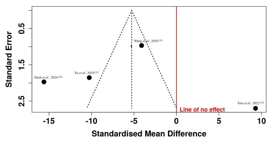
Figure 7: Funnel plot assessing publication bias for studies targeting Oxidative Phosphorylation (OXPHOS) pathway in Pancreatic Ductal Adenocarcinoma (PDAC) (n= 41)
Four studies on FAO pathway in PDAC were analyzed with a total of 76 subjects in the Treatment cohort and 67 subjects in the Control cohort, as shown in Figure 8. Based on the analysis performed, there was no statistical difference between the two groups (SMD = -215.42, 95% CI: -616.27 to -185.44, p > 0.05). The test for overall effect did not show a significant effect. Significant heterogeneity was detected p<0.01, I2 =99%), suggesting inconsistent effects across the studies.
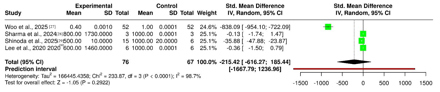
Figure 8: Forest plots comparing mitochondrial metabolic activity between experimental treatment groups and controls in Pancreatic Ductal Adenocarcinoma (PDAC) models across various interventions targeting Fatty acid Oxidation (FAO) pathway in PDAC (n= 76)
Figure 9 presents the funnel plot for studies evaluating FAO-targeting interventions in PDAC. The study estimates show their distribution around the pooled effect across different levels of precision. Visual assessment of the plot did not reveal an obvious asymmetrical pattern suggestive of substantial publication bias.
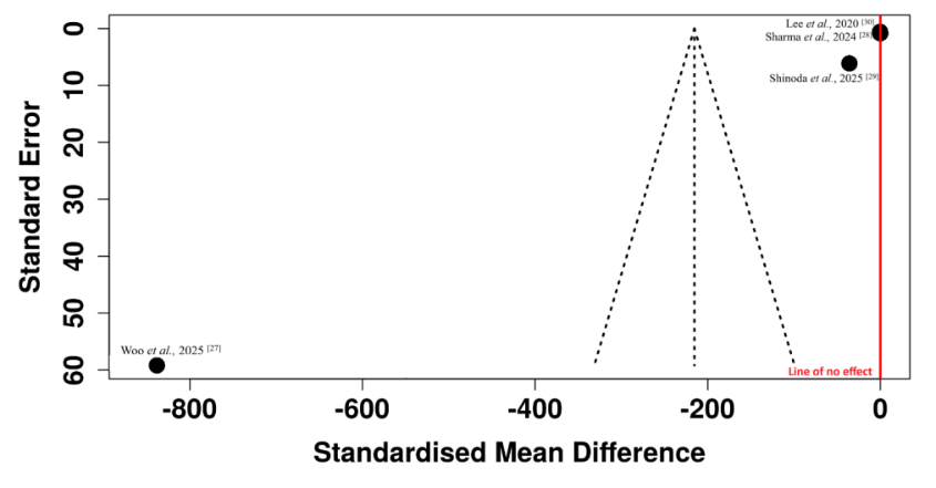
Figure 9: Funnel plot assessing publication bias for studies targeting Fatty acid Oxidation (FAO) pathway in Pancreatic Ductal Adenocarcinoma (PDAC) (n= 76)
Subgroup analyses, including cancer type (AML vs. PDAC) and targeted mitochondrial pathway (OXPHOS vs. FAO), were done to examine possible heterogeneity sources. OXPHOS (four studies) and FAO (three studies) in AML did not have statistically significant differences between the treatment and control groups, with high heterogeneity (I2 = 94.1% and 96.4, respectively). Likewise, subgroup analysis of OXPHOS and FAO in PDAC also showed no important differences (I2 = 95 and 99%, respectively). These subgroup analyses suggested that pooled significance deficiency and the identified heterogeneity were similar in both cancer types and mitochondrial pathways, thus indicating that variability may be attributed to experimental models, interventions, and measurement units. Sensitivity analysis was done by gradual removal of studies to determine their impact on the general effect estimates. The elimination of any of the four groups did not change the direction of the effect estimates or its magnitude significantly, which proved the strength of the primary results. Nevertheless, the continued high heterogeneity among analyses emphasized the variation in study designs and models of xenografts, which suggested that the disparities might be the result of differences in cell lines, xenograft models, or measurement methodologies.
The SYRCLE Risk of Bias Tool was used to evaluate animal xenograft studies as shown in Figure 10. The ROBVIS was used to prepare visualization of the assessment. Overall, the majority of studies demonstrated a moderate ROB. In the animal studies, the majority of the domains presented low risk of baseline similarity, incomplete data on outcomes and selective reporting because outcomes of tumor growth, survival and metabolic assays were objectively measured and reliably reported.
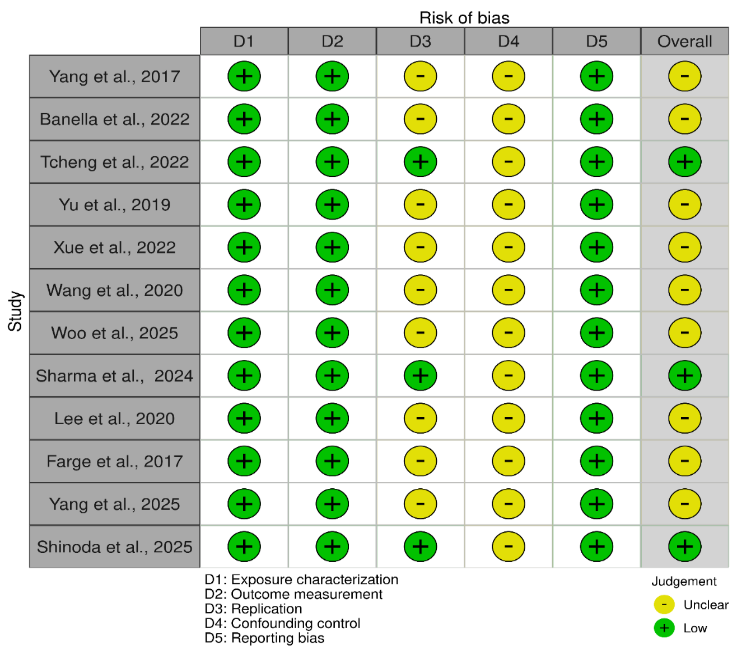
Figure 10: Risk of Bias for In Vitro & Preclinical Experimental Studies using SYRCLE’ s RoB tool (Traffic light plot)
The OHAT Risk of Bias Tool was used to evaluate in-vitro mechanistic studies, as shown in Figure 11. The in vitro studies had low ROB exposure and outcome measures with clear drug treatments and standardized assays. Replication and confounding control had moderate risk because of incomplete reporting, and overall evidence certainty was moderate because of lack of randomization, blinding, and replicates in some studies.
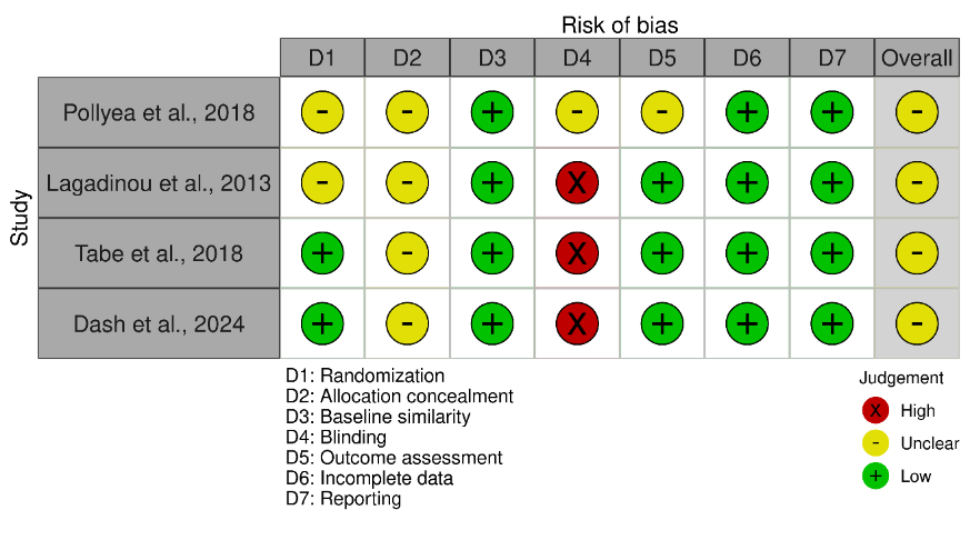
Figure 11: Risk of Bias for In Vitro Mechanistic Studies using OHAT RoB tool (Traffic light plot)
Overall, the quantitative synthesis did not demonstrate statistically significant differences following OXPHOS or FAO pathway targeting in either AML or PDAC. However, substantial heterogeneity was observed across the pooled analyses, indicating considerable variation among the included studies. Funnel plot assessment did not reveal obvious asymmetry suggestive of substantial publication bias. These findings provide a quantitative summary of the available evidence while supporting cautious interpretation of the pooled effects.
The present systematic review and meta-analysis demonstrated that both OXPHOS and FAO are critical mitochondrial metabolic roles in AML and PDAC. The included studies consistently demonstrated that OXPHOS pharmacological or genetic inhibition lowered the respiration rate, ATP production, and the survival of cancer cell lines, especially in cell populations resistant to therapy such as leukemic stem cells. Similarly, the blocking of FAO inhibited the generation of energy in mitochondria and inhibited the growth of tumor cells in AML and PDAC models. Nevertheless, even with the similarly mechanistic tendencies that are observed in each of the aforementioned studies, the combined meta-analysis failed to find any statistically significant differences between treatment and control groups in either of the two types of cancers with regard to OXPHOS or FAO inhibition. This nonsignificant result was supported by a high level of heterogeneity between analyses, which revealed experimental model, intervention, and metabolic measure heterogeneity. However, the general body of evidence corroborated the idea that the hybrid metabolic dependency of cancer cells on both mitochondrial respiration and lipid oxidation was apparent, highlighting the importance of targeting multiple mitochondrial pathways simultaneously to disrupt tumor bioenergetics.
These findings were consistent with emerging literature that showed that mitochondrial metabolism was a key factor in the development of cancer, as well as resistance to therapy. Research showed that LSCs in AML were highly dependent on mitochondrial respiration but not glycolysis 31. In hematologic malignancies, FAO served as a critical catabolic pathway that fueled OXPHOS, particularly in chemotherapy-resistant AML cells 32. In PDAC, tumor cells utilize lipid metabolism and mitochondrial oxidative metabolism to acclimate to nutrient-deprived and hypoxic tumor microenvironments 33. Equally, FAO has been found to be a significant metabolic route that facilitated survival and resistance to chemotherapy in hematologic malignancies 34. Several studies have reported improvements in mitochondrial oxidative metabolism and lipid use in PDAC to allow tumor cells to acclimatize to nutrient-deprived tumor microenvironment 35,36. Recent studies have also implied that metabolic plasticity enabled tumor cells to alternate between glycolysis 37,38, OXPHOS, and FAO in reaction to therapeutic stress, supporting the idea of metabolic plasticity as a contributor to cancer survival 39,40.
Several limitations of the present systematic review should be acknowledged. The pool of studies that could be analyzed by quantitative synthesis was rather limited. Secondly, heterogeneity was found to be substantial among studies because they differed in terms of experimental models, such as cell lines in-vitro, animal xenografts, and translational samples, and they varied concerning metabolism endpoints and measurement units. Third, a number of studies presented their results in terms of various biochemical indicators of mitochondrial activity, which had to be standardized and could have led to variability of pooled effects estimates.
The included studies also had several methodological limitations. The majority of studies were preclinical experimental studies, and only a few of them included clinical or patient-derived samples, which limited the number of studies in direct clinical applications. Also, risk-of-bias assessment noted that a number of studies failed to explicitly report the method of randomization, allocation concealment, and blinding, especially in animal studies. In vitro experiments also did not frequently describe biological replicates and experimental controls for variability in detail.
In the future, studies should adopt translational and clinical studies and research studies to determine the effectiveness of the mitochondrial metabolic inhibitors through effective clinical trials. Specifically, metabolic plasticity can be overcome by simultaneous combination therapeutic approaches targeting both OXPHOS and FAO pathways to decrease the risk of resistance to treatment. A possible solution to enhance the results in metabolically flexible malignancies like AML and PDAC is to combine metabolic targeting with contemporary chemotherapy, immunotherapy, or targeted molecular therapy.
This systematic review and meta-analysis suggested that OXPHOS and FAO contribute to mitochondrial metabolic activity in AML and PDAC under experimental conditions. Inhibition of these pathways was associated with reduced ATP production, cell viability, and tumor growth in the included studies, supporting their potential as therapeutic targets. However, the lack of statistically significant meta-analytic findings, together with substantial heterogeneity and limited sample sizes, warranted cautious interpretation. Further well-designed translational and clinical studies are needed to evaluate combined targeting of OXPHOS and FAO and to identify metabolic biomarkers that may support personalized therapeutic approaches.

Journal of Biomolecules, Pathogenesis and Therapeutics, J Biomol Pathog Ther. 2026;2(2), p69-71 (jbptjournal.org) © 2026 Authors. This work is published by Multidisciplinary Scholarly Advancement and Research MSAR Institute. The full terms of Journal Publishing policy is available at https://jbptjournal.org/index.php/jbpt/-publishing-license and incorporate the Creative Commons Attribution – Non Commercial (CC BY, NC 4.0) License https://creativecommons.org/licenses/by-nc/4.0/. By accessing the work you hereby accept the Terms. Non-commercial uses of the work are permitted without any further permission, provided the work is properly attributed. Publisher’s Note: MSAR Institute remains neutral with regard to jurisdictional claims in published maps and institutional affiliations, and assumes no liability for the scientific accuracy or clinical efficacy of the content herein, as they rest entirely with the authors.